Clinical Summary
An 82-year-old woman was rushed to the emergency room after a fall at home, with slurred speech and sudden right-sided weakness. What looked, at first glance, like a single stroke event turned out to be something more layered: several fresh clots in different parts of the brain, a heart working under strain, and a long-standing habit of chewing tobacco that had never been flagged as a risk factor. This case is a reminder that in elderly patients, stroke rarely has just one cause, and the workup has to dig for all of them.
Patient Presentation
The patient, an 82-year-old woman with no previously documented major illnesses, was brought to the emergency department after a fall at home. Her family reported that she had felt dizzy, then fell near her bed, and immediately afterward her speech became slurred and her right arm and leg felt weak, with the right side of her face appearing to droop. There was no prior history of stroke, heart disease, or diabetes on record, though on direct questioning the family confirmed a long-standing habit of chewing smokeless tobacco (a form of tobacco use, distinct from smoking, that is still a recognised cardiovascular and cerebrovascular risk factor).
On arrival, she was conscious and oriented but had visible bruising over her right knee and forearm from the fall.
Clinical Examination
- Blood pressure: 192/86 mmHg (significantly elevated)
- Pulse: 68/min, Temperature: 97.2°F, Oxygen saturation: 96% on room air
- Random blood sugar: 148 mg/dL
- Neurological findings: Right-sided facial weakness, right upper and lower limb power reduced to 3/5 (on a scale where 5/5 is full strength)
- Systemic exam: Chest clear, abdomen soft and non-tender, heart sounds normal
- NIH Stroke Scale: 7, indicating a moderate deficit at presentation
Investigations and Findings
Brain imaging (NCCT then MRI Brain)
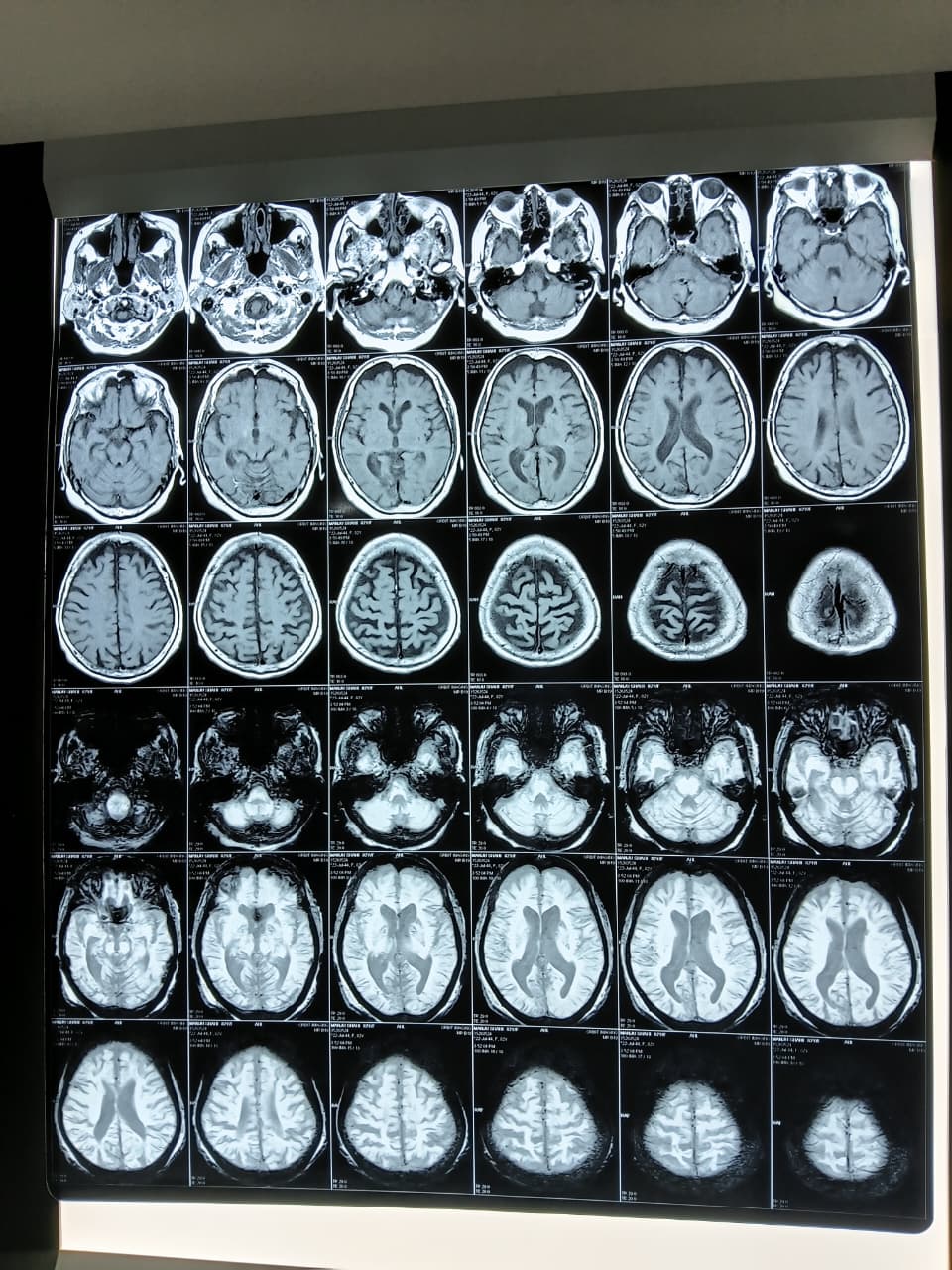
- A non-contrast CT (NCCT) head done immediately on arrival showed an old lacunar infarct (a small, deep stroke from years past) with no bleeding, which cleared her for stroke-specific treatment.
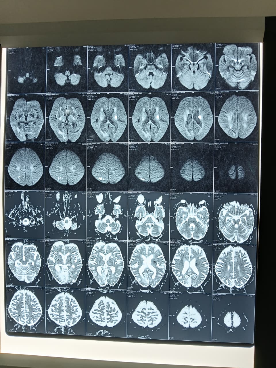
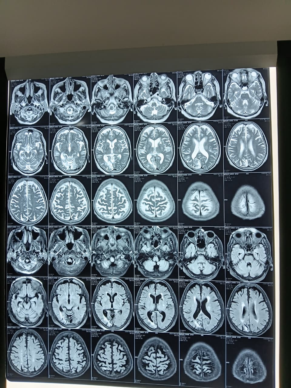
- MRI Brain with diffusion sequences then revealed the fuller picture:
- An acute infarct in the left centrum semiovale and corona radiata (deep white matter pathways carrying signals to and from the brain's surface), about 1.6 x 1.0 cm.
- Additional small acute infarcts in the right high fronto-parietal region, the largest about 1.9 x 0.9 cm, described as likely ischaemic/embolic (caused by a clot travelling from elsewhere in the body, rather than local narrowing of a single vessel).
- An older, partially gliotic (scarred) lesion in the right occipital region, representing a prior, unrecognised stroke.
- A small gliotic lacunar infarct in the left ganglio-capsular region, again old.
- Diffuse age-related cerebral and cerebellar atrophy with chronic small-vessel ischaemic changes, common in this age group.
The presence of acute infarcts in more than one vascular territory at the same time is an important clue. It shifts the differential away from a single blocked artery and toward a source that can shower clots to multiple places at once, most often the heart or a diseased major vessel.
Cardiac evaluation (2D Echo)
- LVEF (left ventricular ejection fraction): approximately 45%, mildly reduced (the heart's pumping efficiency was below the normal 55-65% range)
- Regional wall motion abnormality present, with hypokinesia (reduced movement) of the mid posterior, basal lateral, and basal inferior walls
- Mild concentric LV hypertrophy (thickening of the heart muscle, typically from long-standing high blood pressure)
- Raised left ventricular filling pressure, mild mitral regurgitation, mild aortic regurgitation, thickened aortic valve, mild tricuspid regurgitation
- No intracardiac clot, vegetation, or pulmonary embolism seen
Follow-up ECG (done after discharge) showed a bifascicular block (a conduction abnormality in the heart's electrical wiring, involving two of its three main pathways), adding a structural and electrical dimension to the cardiac picture.
Laboratory work-up
- Vitamin B12: markedly elevated (>2000 pg/mL)
- HbA1c: 5.5% (non-diabetic range), estimated average glucose 111 mg/dL
- Liver function: essentially preserved, with a mildly raised alkaline phosphatase
- Kidney function, electrolytes, and complete blood count: within acceptable limits for age
- ESR: 40 mm/hr, mildly elevated
Serum homocysteine testing and an overnight EEG were advised as part of a complete stroke work-up, but after detailed counselling regarding the risks and benefits, the family chose to decline both, and this refusal was documented in writing.
Diagnosis
Final diagnosis: Acute ischaemic stroke with multiple acute infarcts involving the right fronto-parietal region and left anterior ganglio-capsular region, superimposed on old lacunar and gliotic changes, with newly diagnosed accelerated hypertension.
The combination of infarcts in more than one arterial territory, a heart with regional wall motion abnormality and mildly reduced ejection fraction, and a newly identified conduction defect (bifascicular block) raised suspicion for a cardioembolic contribution (clots forming in a weakened or irregularly beating heart and travelling to the brain), alongside the more typical small-vessel disease expected at this age. A single-artery, atherosclerotic explanation was considered less likely given the multi-territory pattern, though full etiological workup (carotid Doppler was performed; further arrhythmia monitoring was advised as an outpatient) remained ongoing at the time of discharge. The dramatically elevated blood pressure, with no prior diagnosis or treatment, was classified as accelerated hypertension and treated as a second, independent driver of her vascular risk.
Treatment Approach
She was managed as an acute stroke admission with:
- Antiplatelet therapy (aspirin, later combined briefly with clopidogrel, which was discontinued at follow-up once the cardiac picture was clarified)
- High-intensity statin therapy for plaque stabilisation and lipid control
- Neuroprotective and metabolic support with citicoline-piracetam and S-adenosyl-L-methionine with ginkgo biloba combinations
- Antihypertensive optimisation using a telmisartan, cilnidipine, and chlorthalidone combination, later supplemented with metoprolol succinate for both blood pressure and heart rate control given the cardiac findings
- Betahistine for her dizziness symptoms
- Advice for physiotherapy for the residual weakness, and BP monitoring twice daily at home
At the follow-up visit, with the ECG and echo findings reviewed, clopidogrel was stopped and cardiology recommended no active cardiac intervention was needed at that time, with continued medical management and monitoring of NT-proBNP as an outpatient.
Outcome and Follow-Up
The patient remained hemodynamically stable throughout admission. Her blood pressure improved with treatment, though her right-sided weakness had progressed slightly during the stay before stabilising, and she was discharged with residual right hemiparesis (weakness on one side of the body), right upper limb power 2/5 and right lower limb power 3/5. She was discharged in stable condition with clear instructions for strict medication adherence, a target blood pressure of 130/80 mmHg, LDL cholesterol target below 70 mg/dL, complete avoidance of smoking, alcohol, and tobacco, and aggressive physiotherapy and rehabilitation. Follow-up was arranged in both neurology and cardiology outpatient clinics within a week, with periodic lipid profile and renal function monitoring given the new antihypertensive regimen.
Clinical Pearls and Teaching Points
For Clinicians
- Multi-territory acute infarcts on MRI, especially spanning both anterior circulation hemispheres, should always prompt a focused search for an embolic source, cardiac or otherwise, rather than being filed away as "generalised small-vessel disease of the elderly."
- A mildly reduced LVEF (around 45%) with regional wall motion abnormality in a patient without a known prior cardiac event deserves rhythm monitoring; a bifascicular block found incidentally on a routine ECG can be the first visible clue to an underlying conduction system disease that predisposes to arrhythmia-related cardioembolism.
- Newly diagnosed, markedly elevated blood pressure at the time of stroke (accelerated hypertension) should be treated as a distinct, independently modifiable risk factor, not simply a reactive rise from the acute event; it needs its own long-term target and monitoring plan.
- Smokeless tobacco use is frequently under-asked and under-documented in elderly patients, particularly women, yet it carries recognised cardiovascular and cerebrovascular risk; a specific, direct question about chewing tobacco should be part of every stroke history.
- When families decline recommended investigations (such as overnight EEG or homocysteine testing) after full counselling, thorough documentation of informed refusal is essential, both for patient safety and medico-legal clarity, without compromising the rest of the care plan.
For Patients and Families
- A stroke in an elderly relative is rarely caused by just one thing. It is common for doctors to order several tests, brain imaging, heart imaging, blood work, because more than one factor is often working together.
- High blood pressure can exist silently for years without symptoms. A stroke can sometimes be the first sign that it was there all along, which is why regular BP checks matter even without complaints.
- Chewing tobacco is not a "safer" alternative to smoking. It carries real risks for the heart and brain and should be discussed openly with your doctor, even if it feels like a minor habit.
- If your doctor recommends a test and you decide against it, make sure you understand exactly what that decision means for future risk, and keep the door open to revisit it later if you change your mind.
This case was managed by Dr. Bhupesh Kumar Mansukhani, MBBS (Australia), MD (Medicine), DM (Neurology), Fellow in Stroke Medicine and Advanced Neurological Disorders (Harvard Medical School), Neurologist and Director, NeuroMet Wellness Care and Diagnostics, Gurgaon. For appointments and consultations, visit neurometwellness.com/booking.php
Disclaimer: Patient details have been de-identified and shared with appropriate consent. This case study is for educational purposes only and does not constitute medical advice.